A Roman-period cranial tumor case from Sima de Marcenejas
The CENIEH publishes a study of a case of meningioma in a Roman-period cranium. Using micro-computed tomography, it was possible to obtain hundreds of X-ray images to create a 3D model and visualize the interior of the cranium in detail.

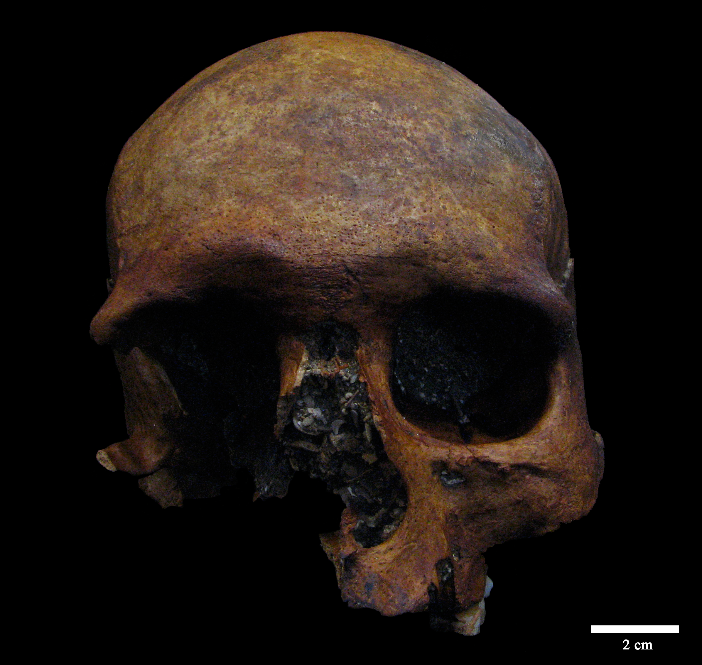
A multidisciplinary team at the Centro Nacional de Investigación sobre la Evolución Humana (CENIEH) has published a paper in the journal Virtual Archaeology Review on a Roman-period meningioma (cranial tumor) in the Iberian Peninsula. The finding of this skull, together with signs of cranial lesions in the same individual, offers new data about the health of past populations.
The cranium was discovered during a caving expedition to the Sima de Marcenejas, in Lastras de Teza (Burgos), thanks to the collaboration of the caving groups Gaem, Takomano, Geoda, Flash, and A.E.Get, which played a fundamental role in its recovery.
It was then carried to the CENIEH, where it was subjected to a meticulous process by the team at the Conservation and Restoration Laboratory. After all this work, it was possible to identify it as belonging to an adult male individual who lives in the final centuries of the Roman Empire.
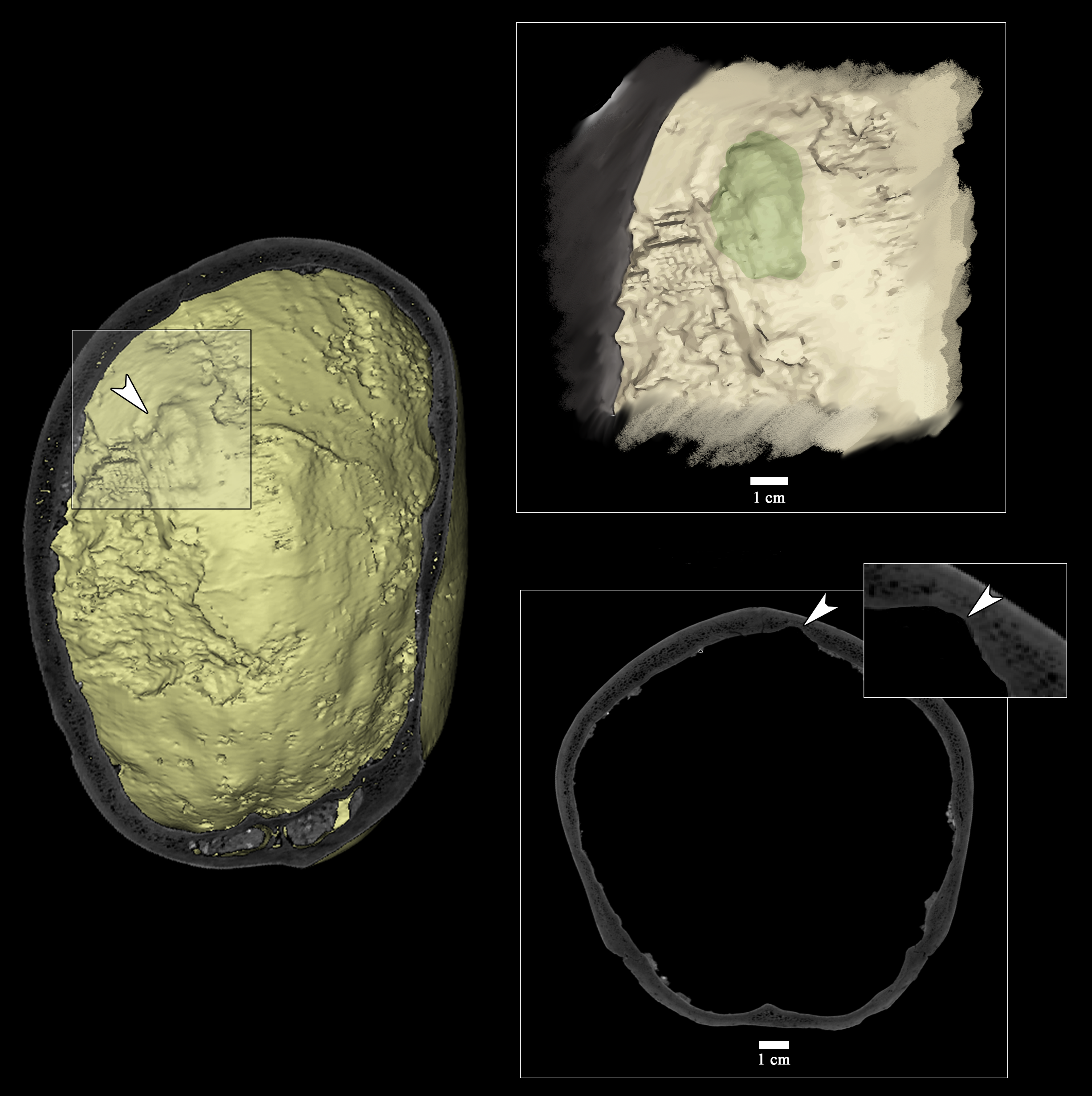
The main objective of this investigation was to understand the possible diseases that affected this person during life. Cutting-edge techniques such as micro-computed tomography (MicroCT) were used to do this, making it possible to obtain hundreds of X-ray images to create a 3D model and visualize the interior of the cranium in detail.
Virtual autopsy
Essentially, micro-computed tomography allows conducting a sort of “virtual autopsy” on the individual, and this revealed the presence of four cranial lesions, all of antemortem origin, that is, injuries showing evidence of curing processes, indicating that they happened while the individual was alive.
Of the four lesions identified, three of them were on the outside of the skull, and show evidence compatible with injuries produced intentionally. This is because they are on the top of the head, which is not typical for lesions caused by accidents such as falls.
Moreover, two of them show characteristics consistent with wounds inflicted by sharp and blunt objects. This raises the possibility that they were the result of violent attempts against this individual’s life.
Intracranial lesion
The fourth lesion is inside the skull. It is characterized by being a depression of circular morphology which had eliminated part of the bone holding it.
After studying the characteristics of the lesion and conducting a comparative analysis with different pathologies such as infections, metabolic or genetic diseases, or a neoplasia, the conclusion was reached that it was probably caused by a tumor inside the skull, a possible meningioma. This meningioma is the first case of this condition for these chronologies in the Iberian Peninsula, which is a region with few records of these tumors from antiquity.
“What is interesting about this finding is that it offers a window onto the health of past populations, and raises fundamental questions for us about the ability of individuals to survive these conditions, and their quality of life thereafter”, says Daniel Rodríguez-Iglesias, lead author of this paper.

Press release from Centro Nacional de Investigación sobre la Evolución Humana – CENIEH


